
Flat Feet in Adults: When Should You Be Concerned?
 webmaster on
 Flexible flat feet in adults are common, often asymptomatic, and usually require only education, supportive footwear, and activity modification. Concern arises with persistent or progressive medial ankle or arch pain, activity-limiting symptoms, recurrent ankle sprains your mobile podiatrist, new stiffness or loss of arch flexibility, rapid collapse (suggesting neuropathic/Charcot change), or systemic/neurologic signs. Evaluation includes focused history, exam, weightbearing radiographs and targeted imaging. Further information outlines causes, diagnostic steps, and treatment thresholds.
When Flat Feet Are Normal and When They’re a Problem
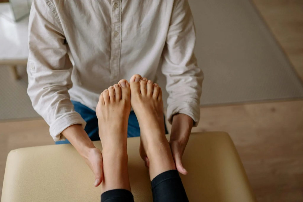
When evaluating adult flat feet, clinicians distinguish flexible, asymptomatic flattening—common and often benign—from rigid deformities or cases accompanied by pain, dysfunction https://peoplepodiatry.com.au/, or progressive change that indicate pathology.
Examiners assess whether the presentation reflects developmental variations persisting into adulthood or acquired collapse. Flexible deformities demonstrate functional flexibility with normal arch return on toe rise and nonpainful gait; these often require no intervention beyond education, supportive footwear, and activity modification.
In contrast, loss of flexibility, progressive valgus alignment, tendon dysfunction, or new mechanical symptoms prompt imaging and targeted treatment.
Objective assessment includes weightbearing alignment, single-leg heel rise, and comparative limb evaluation to preserve mobility and autonomy.
Management prioritizes restoring function and preventing decline while respecting patient goals for activity and independence.
Key Symptoms That Suggest an Underlying Condition
Recognize symptoms that raise concern for pathology rather than benign flexible flatfoot: persistent or progressive medial ankle and hindfoot pain, activity-limiting plantar or arch pain, lateral forefoot overload, recurrent ankle sprains, progressive hindfoot valgus, and new onset stiffness or loss of arch flexibility. Such features warrant prompt evaluation.
Severe pain disproportionate to activity, sudden swelling after minimal trauma, or nocturnal pain suggest structural compromise or inflammatory disease and merit imaging and specialist referral.
Functional decline—reduced walking tolerance, gait instability, or inability to resume preferred activities—indicates progressive dysfunction.
Neurologic signs (numbness, weakness) or systemic symptoms (fever, unexplained weight loss) broaden the differential and require expedited assessment.
Early identification preserves mobility and autonomy; clinical judgment should guide timely investigation.
Common Causes in Adults: PTTD, Arthritis, Nerve and Tendon Issues
Among adults, acquired flatfoot most commonly reflects mechanical or inflammatory disruption of key supporting structures, especially posterior tibial tendon dysfunction (PTTD), degenerative or inflammatory arthritis of the hindfoot and midfoot, and neuropathic or tendinous disorders that alter load distribution.
Adult acquired deformity often begins with Posterior tibialis failure—attenuation, tearing, or degeneration—producing progressive valgus alignment and midfoot collapse.
Arthritis (osteoarthritis, post-traumatic, rheumatoid) erodes joint congruity and passive support, accelerating deformity and pain.
Neuropathic collapse from diabetic neuropathy or Charcot arthropathy results in rapid architecture loss with sensory loss and joint destruction.
Other tendon dysfunctions (peroneals, Achilles) and iatrogenic factors modify biomechanics.
Management priorities include gait preservation, risk reduction for ulceration or progression, and targeted interventions to restore stable, pain-free function.
How Doctors Diagnose Flat Feet and What Tests to Expect
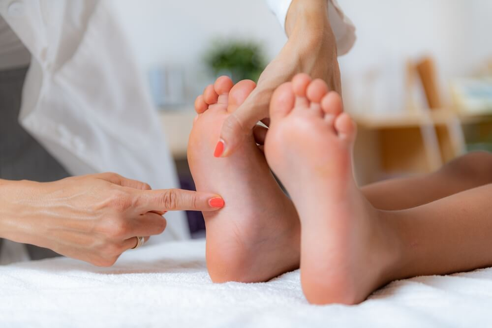
Clinicians evaluate adult flatfoot through a structured clinical history and targeted physical examination to determine etiology, severity, and functional impact. Assessment documents pain, onset, prior injury, systemic disease, and activity limitations.
Physical tests include inspection of arch height, heel alignment, single-leg heel rise, and range of motion at subtalar and ankle joints. Objective evaluation often uses gait analysis to quantify dynamic pronation, stride asymmetry, and compensatory patterns.
Imaging begins with weight bearing Xray series to assess alignment, joint space, and osseous deformity; oblique and hindfoot views may be added. Advanced imaging (CT or MRI) is reserved for suspected arthritis, tendon pathology, or complex deformity.
Diagnosis integrates history, exam, dynamic analysis, and imaging to guide appropriate management decisions.
Treatment Options and When Surgery Becomes Necessary
In managing adult flatfoot, treatment begins with a graded approach that matches interventions to severity, symptoms, and underlying pathology. Initial care emphasizes nonoperative measures: activity modification, anti-inflammatory medication, targeted Physical therapy to restore strength and proprioception, and Shoe modifications including supportive footwear and custom orthoses to redistribute load. Weight management and gradual return to activity support sustained improvement.
Indications for surgery include persistent pain despite optimized conservative care, progressive deformity, or neuromuscular compromise affecting function. Surgical options are pathology-specific: tendon reconstruction, osteotomy, arthrodesis, or combined procedures to realign and stabilize the medial column. Decision-making should integrate imaging, functional assessment, patient goals for mobility and independence, and realistic expectations about recovery and outcomes.
Conclusion
Flat feet in adults often reflect benign anatomic variance, but persistent pain, progressive deformity, functional decline or neurologic signs warrant evaluation. Clinical assessment supplemented by weight‑bearing imaging and targeted tests distinguishes flexible, asymptomatic pes planus from posterior tibial tendon dysfunction, inflammatory or degenerative arthropathy, neuropathy or traumatic causes. Initial management is conservative and evidence‑based—activity modification, orthoses, physiotherapy and pharmacologic measures—while surgery is reserved for refractory pain, instability or structural collapse confirmed by objective findings.